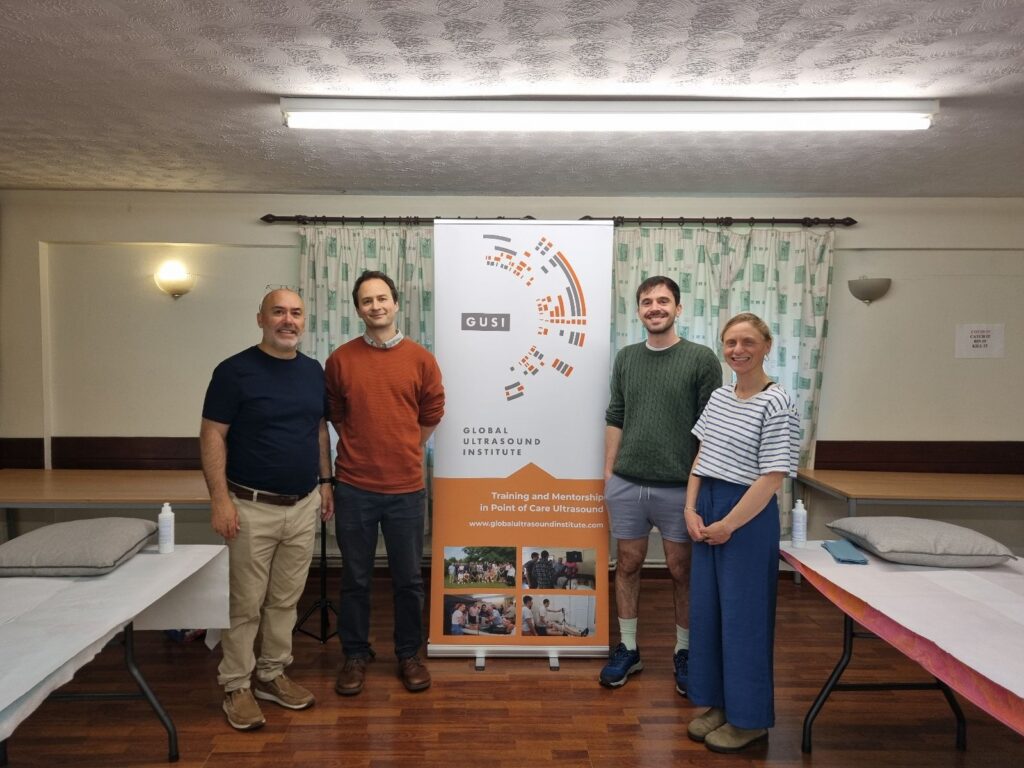
As part of the Global Ultrasound Institute’s mission to empower healthcare providers worldwide with point-of-care ultrasound (POCUS) skills , three GUSI Fellows in the United Kingdom recently participated in an intensive hands-on ultrasound training workshop.
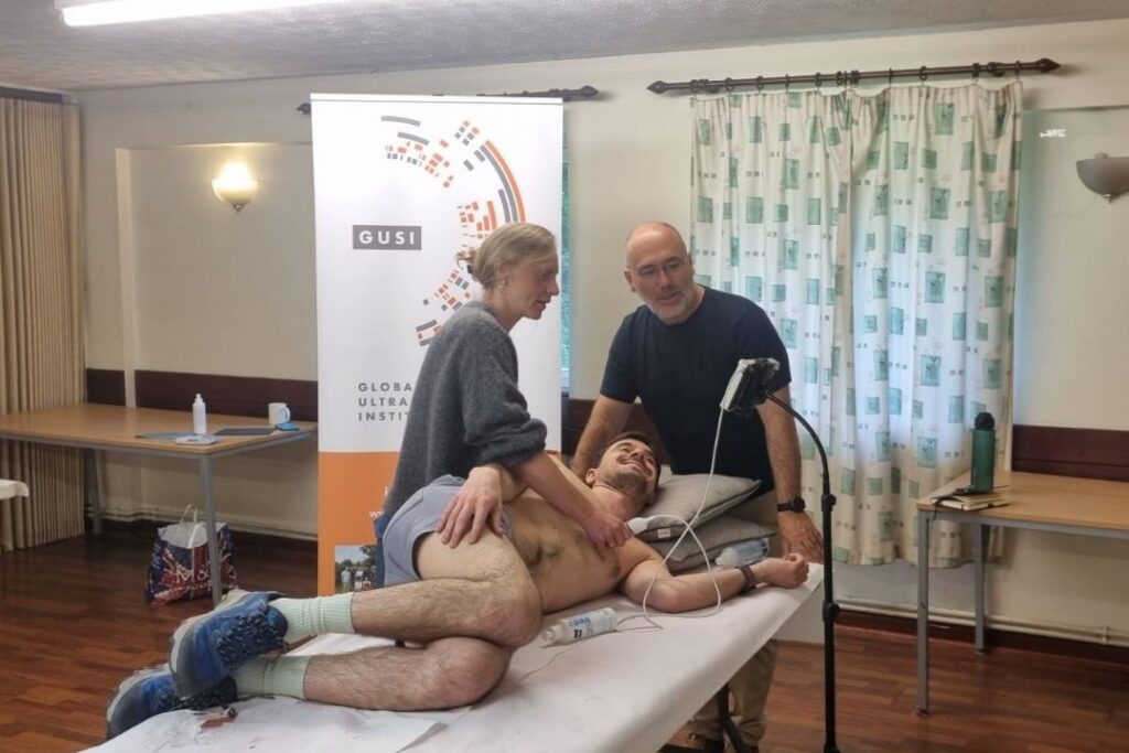
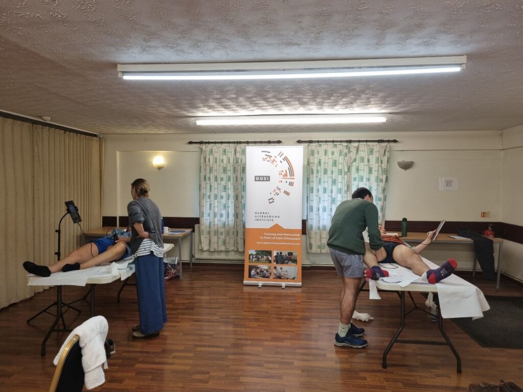
Dr. Viviano, Dr. Averill, and Dr. Mannings – family physicians from Scotland, Devon, and Cornwall – completed a 6-hour POCUS training session under the expert guidance of Dr. Pascual Daza-Ramirez (GUSI’s Education Director for the UK & Europe ). This immersive experience was designed to enhance their clinical ultrasound proficiency through real patient scanning and immediate expert feedback. Such professional medical education in POCUS is increasingly crucial for primary care and family medicine, where rapid, bedside diagnostic insights can greatly improve patient care .
Expert Mentorship: A Key to POCUS Mastery

Learning ultrasound is a hands-on skill, and having an experienced mentor significantly accelerates the learning curve. Throughout the session, Dr. Pascual Daza-Ramirez provided one-on-one instruction, demonstrating proper probe techniques, optimal ultrasound windows, and interpretation of findings. His expertise as a senior POCUS educator was invaluable – guiding the fellows to recognize anatomy and pathology in real time. Effective POCUS use is highly operator-dependent, so this kind of supervised training helps clinicians avoid common pitfalls and build confidence . Under Dr. Daza-Ramirez’s tutelage, the fellows gained practical experience in scanning and diagnosing, reinforcing concepts from their online studies with live patients. By the end of the day, they had not only honed their technical scanning skills but also learned clinical pearls – for example, how to adjust imaging depth for an abdominal aorta exam or how to spot subtle B-lines in a lung scan – that will inform their everyday practice.
Scanning Key Anatomical Structures with Confidence
Under direct supervision, the GUSI Fellows practiced scanning a broad range of key anatomical structures commonly evaluated in primary care POCUS, including:
- Heart – Focused cardiac ultrasound to visualize chambers and function (e.g. detecting pericardial effusions or reduced ejection fraction).
- Lungs – Thoracic ultrasound to assess lung sliding, B-lines, and pleural effusions (useful for diagnosing pneumonia or heart failure).
- Kidneys – Renal scans to check for hydronephrosis or kidney stones (ruling out obstructive uropathy in patients with flank pain).
- Gallbladder & Liver – Abdominal RUQ scans to identify gallstones, cholecystitis signs (Murphy’s sign) or free fluid around the liver (as in trauma assessments).
- Abdominal Aorta – Ultrasound screening for abdominal aortic aneurysms (AAA), measuring aortic diameter to catch life-threatening enlargement early.
- Pelvic Organs – Pelvic ultrasound to evaluate bladder volume or, in appropriate cases, confirm intrauterine pregnancy (which effectively rules out ectopic pregnancy in early obstetrics scenarios).
- Femoral Veins – Compression ultrasound of the femoral vein (and popliteal vein) to detect deep venous thromboses (DVT) in patients with leg swelling or clot risk.
Practicing these scans on real patients gave the fellows valuable hands-on repetition in image acquisition and interpretation. Notably, these POCUS applications cover many high-impact diagnoses in outpatient and acute care. POCUS has been shown to have excellent accuracy for detecting conditions like AAA, DVT, gallbladder disease, pneumonia, and heart failure when performed by trained clinicians . By mastering cardiac, lung, abdominal, pelvic, and vascular ultrasound techniques, the fellows are equipping themselves to quickly recognize emergent conditions (such as cardiac tamponade or a ruptured aneurysm) and to enhance routine assessments (such as distinguishing cellulitis from abscess, or confirming a consolatory pneumonia) right at the bedside. This breadth of scanning practice reflects a comprehensive POCUS skill set that is increasingly expected of modern primary care and family medicine practitioners.

Scanning Key Anatomical Structures with Confidence
The Sonoscanner U-Lite, an ultraportable high-performance ultrasound device used during the training, offers high-definition imaging in a pocket-sized form factor .
A huge thanks is owed to Sonoscanner for providing the U-Lite device to power this hands-on training. The Sonoscanner U-Lite is a handheld ultrasound unit known for its exceptional image quality and nearly instant boot-up time . Weighing only around 600–700 grams and featuring a 7-inch high-resolution touchscreen, the U-Lite gave the fellows the freedom to scan anywhere in the clinic without being tethered to a full-size machine. Its portability and ease of use meant more scanning time and less waiting, which is ideal for fast-paced teaching sessions. During the 6-hour workshop, the device performed impressively – from cardiac apical views to abdominal deep scans – demonstrating how modern ultraportable ultrasound technology can bring imaging directly to the point of care. By training with the U-Lite, the fellows also became familiar with a device they can realistically use in their own practice settings (rural clinics or community hospitals), where quick deployment of ultrasound can make a critical difference. This synergy of skilled instruction and cutting-edge equipment exemplifies how #UltrasoundEducation is evolving, making POCUS training more accessible and impactful than ever.
Extending Learning with ScanHub™ and ScanOne™
One unique angle of GUSI’s educational approach is the integration of digital tools like ScanHub™ and ScanOne™ to complement in-person training. After the workshop, the fellows can continue to refine their skills using GUSI’s online platforms. ScanHub is GUSI’s comprehensive POCUS learning ecosystem – included with every GUSI membership – which provides access to online course materials, a library of ultrasound videos and cases, knowledge quizzes, and even an AI tutor (“Sage AI”) for on-demand questions . Through ScanHub’s ScanFolio archive, the fellows can upload and store their own ultrasound clips securely, track their scanning performance, and monitor their progress via personalized dashboards . This creates a feedback loop where skills learned in the hands-on session are reinforced by reviewing images and identifying areas for improvement over time.
Meanwhile, ScanOne is GUSI’s new cloud-based tool specifically designed for POCUS learners to get expert feedback on their scans. Using ScanOne, the fellows can upload the ultrasound images or clips they acquire in their day-to-day practice and request feedback or quality assurance from GUSI’s faculty – all within a secure, educational platform . This means that even after the UK training event, Dr. Viviano, Dr. Averill, and Dr. Mannings can perform scans on their own patients back home and have an expert mentor virtually “over-the-shoulder” to review those images. ScanOne is strictly for education (not clinical diagnosis), but it effectively bridges the gap between training and practice by allowing continuous mentorship and learning opportunities . The combination of these platforms – the ScanHub learning suite and the ScanOne feedback tool – ensures that the fellows’ growth doesn’t stop when the workshop ends. It exemplifies how modern medical education leverages technology to create a blended learning experience: hands-on practice supported by ongoing digital mentorship and resources.

POCUS: Transforming Practice for UK Family Physicians
By the conclusion of the 6-hour session, the GUSI Fellows had dramatically sharpened their ultrasound skills, and more importantly, they gained the confidence to apply POCUS in their everyday clinical decision-making. This training is a significant step in bringing advanced diagnostic capability to primary care settings across the UK. Equipped with their new scanning proficiency and supported by GUSI’s online tools, these doctors are poised to implement POCUS for the benefit of their patients – whether it’s swiftly diagnosing heart failure in a rural Scotland clinic, detecting a hidden pneumonia in Devon, or ruling out a DVT in a Cornwall community hospital. The hands-on experience under expert guidance has prepared them to perform focused exams that can expedite diagnoses and triage appropriately, which ultimately improves patient outcomes and saves lives .


